


Knee
Normal Anatomy of the Knee Joint
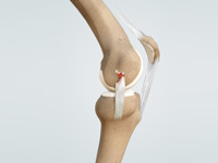
The knee is made up of four bones. The femur or thighbone is the bone connecting the hip to the knee. The tibia or shinbone connects the knee to the ankle. The patella (kneecap) is the small bone in front of the knee and rides on the knee joint as the knee bends. The fibula is a shorter and thinner bone running parallel to the tibia on its outside. The joint acts like a hinge but with some rotation.
The knee is a synovial joint, which means it is lined by synovium. The synovium produces fluid lubricating and nourishing the inside of the joint. Articular cartilage is the smooth surfaces at the end of the femur and tibia. It is the damage to this surface which causes arthritis.
Femur
The femur (thighbone) is the largest and the strongest bone in the body. It is the weight bearing bone of the thigh. It provides attachment to most of the muscles of the knee.
Condyle
The two femoral condyles make up for the rounded end of the femur. Its smooth articular surface allows the femur to move easily over the tibial (shinbone) meniscus.
Tibia
The tibia (shinbone), the second largest bone in the body, is the weight bearing bone of the leg. The menisci incompletely cover the superior surface of the tibia where it articulates with the femur. The menisci act as shock absorbers, protecting the articular surface of the tibia as well as assisting in rotation of the knee.
Fibula
The fibula, although not a weight bearing bone, provides attachment sites for the Lateral collateral ligaments (LCL) and the biceps femoris tendon.
The articulation of the tibia and fibula also allows a slight degree of movement, providing an element of flexibility in response to the actions of muscles attaching to the fibula.
Patella
The patella (kneecap), attached to the quadriceps tendon above and the patellar ligament below, rests against the anterior articular surface of the lower end of the femur and protects the knee joint. The patella acts as a fulcrum for the quadriceps by holding the quadriceps tendon off the lower end of the femur.
Menisci
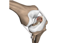
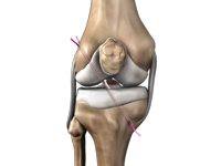
The medial and the lateral meniscus are thin C-shaped layers of fibrocartilage, incompletely covering the surface of the tibia where it articulates with the femur. The majority of the meniscus has no blood supply and for that reason, when damaged, the meniscus is unable to undergo the normal healing process that occurs in the rest of the body. The menisci act as shock absorbers, protecting the articular surface of the tibia as well as assisting in rotation of the knee. As secondary stabilizers, the intact menisci interact with the stabilizing function of the ligaments and are most effective when the surrounding ligaments are intact.
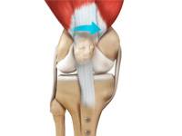
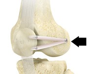
Anterior Cruciate Ligament (ACL)
The anterior cruciate ligament (ACL) is the major stabilizing ligament of the knee. The ACL is located in the center of the knee joint and runs from the femur (thigh bone) to the tibia (shin bone), through the center of the knee. The ACL prevents the femur from sliding backwards on the tibia (or the tibia sliding forwards on the femur). Together with the posterior cruciate ligament (PCL), ACL stabilizes the knee in a rotational fashion. Thus, if one of these ligaments is significantly damaged, the knee will be unstable when planting the foot of the injured extremity and pivoting, causing the knee to buckle and give way.
Posterior Cruciate Ligament (PCL)
Much less research has been done on the posterior cruciate ligament (PCL) because it is injured far less often than the ACL.
The PCL prevents the femur from moving too far forward over the tibia. The PCL is the knee’s basic stabilizer and is almost twice as strong as the ACL. It provides a central axis about which the knee rotates.
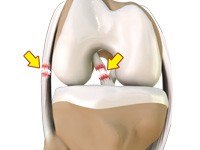
Collateral Ligaments
Collateral Ligaments prevent hyperextension, adduction, and abduction
- Superficial MCL (Medial Collateral Ligament) connects the medial epicondyle of the femur to the medial condyle of the tibia and resists valgus force
- Deep MCL (Medial Collateral Ligament) connects the medial epicondyle of the femur with the medial meniscus
- LCL (Lateral Collateral Ligament) entirely separate from the articular capsule, connects the lateral epicondyle of the femur to the head of the fibula and resists varus force
Knee Conditions
Knee Arthritis
Arthritis is a general term covering numerous conditions where the joint surface or cartilage wears out. The joint surface is covered by a smooth articular surface that allows pain free movement in the joint. This surface can wear out for a number of reasons; often the definite cause is not known.
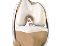
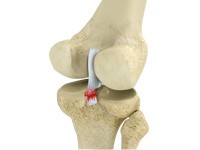
Anterior Cruciate Ligament (ACL) Tear
The anterior cruciate ligament, or ACL, is one of the major ligaments of the knee that is located in the middle of the knee and runs from the femur (thigh bone) to the tibia (shin bone). It prevents the tibia from sliding out in front of the femur. Together with posterior cruciate ligament (PCL) it provides rotational stability to the knee.
Ligament Injury
The knee is a complex joint which consists of bone, cartilage, ligaments and tendons that make joint movements easy and at the same time it is more susceptible to various kinds of injuries. Knee problems may arise if any of these structures get injured by overuse or suddenly during sports activities. Pain, swelling, and stiffness are the common symptoms of any damage or injury to the knee.
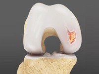
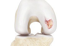
Cartilage Defect / Injury
Articular or hyaline cartilage is the tissue lining the surface of the two bones in the knee joint. Cartilage helps the bones move smoothly against each other and can withstand the weight of the body during activities such as running and jumping. Articular cartilage does not have a direct blood supply to it so has less capacity to repair itself.
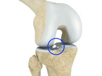
Loose Body
Loose bodies are small loose fragments of cartilage or a bone that float around the joint. The loose bodies can cause pain, swelling, locking and catching of the joint. Loose bodies occur if there is bleeding within the joint, death of tissues lining the joints associated with tuberculosis, osteoarthritis, and rheumatoid arthritis.
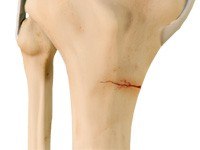
Knee Fracture
A fracture is a condition in which there is break in the continuity of the bone. In younger individuals, these fractures are caused from high energy injuries, as from a motor vehicle accident. In older people the most common cause is weak and fragile bone.
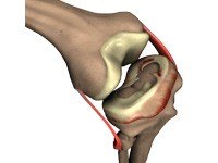
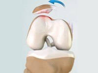
Meniscus Tear
Meniscus tear is the commonest knee injury in athletes, especially those involved in contact sports. A suddenly bend or twist in your knee cause the meniscus to tear. This is a traumatic meniscus tear. Elderly people are more prone to degenerative meniscal tears as the cartilage wears out and weakens with age.
Patellofemoral Instability
The knee can be divided into three compartments: patellofemoral, medial and lateral compartment. The patellofemoral compartment is the compartment in the front of the knee between the knee cap and thigh bone. The medial compartment is the area on the inside portion of the knee, and the lateral compartment is the area on the outside portion of the knee joint.
Knee Procedures
Meniscus Repair
Meniscal transplantation is a surgical procedure to replace the damaged meniscus of the knee with healthy cartilage.The meniscus is a C-shaped cartilage ring that acts like a cushion between the shinbone and the thighbone. Each of your knees has two menisci - one on the inside (medial aspect) and the other on the outside (lateral aspect) of your knee. Apart from the cushioning effect, the menisci also provide stability to the knee.
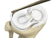
Partial Meniscectomy
Partial meniscectomy is a surgical procedure to remove the torn portion of the meniscus from the knee joint. Meniscus is the C-shaped cartilage located in the knee that lubricates the knee joint, acts as shock-absorber, and controls the flexion and extension of joint. Meniscal tears can occur at any age, but are more common in athletes playing contact sports.
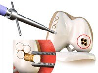
Osteochondral Allograft
An osteochondral allograft is a piece of tissue taken from a diseased donor to replace damaged cartilage that lines the ends of bones in a joint. A section of cartilage and bone is removed, shaped to precisely fit the defect and then transplanted to reconstruct the damage.
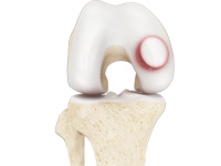
Cartilage Restoration
Articular cartilage is the smooth, shiny, white tissue covering the ends of bones those form a joint. Articular cartilage reduces friction when bones glide over each other, making the movements smooth and painless. It also acts as a shock-absorber to help prevent traumatic injuries to the bones. When cartilage is damaged, it can cause painful movements and limited joint mobility and eventually progress to osteoarthritis.
Patella Stabilization
Patella (knee cap) is a protective bone attached to the quadriceps muscles of the thigh by quadriceps tendon. Patella attaches with the femur bone and forms a patellofemoral joint. Patella is protected by a ligament which secures the kneecap from gliding out and is called as medial patellofemoral ligament (MPFL).
Posterior Cruciate Ligament Reconstruction
Posterior cruciate ligament (PCL), one of four major ligaments of the knee, is situated at the back of the knee. It connects the thighbone (femur) to the shinbone (tibia). The PCL limits the backward movement of the shinbone. PCL injuries are very rare and difficult to detect when compared to other knee ligament injuries.
Posterolateral Corner Reconstruction
Posterolateral corner injury is damage or injury to the structures of the posterolateral corner. The structures of the posterolateral corner include the lateral collateral ligament, the popliteus tendon, and the popliteo-fibular ligament. Injuries to the posterolateral corner most often occur with athletic trauma, motor-vehicle accidents and falls.
Medial Collateral Ligament Repair & Reconstruction
Medial collateral ligament (MCL) is one of four major ligaments of the knee that connects the femur (thigh bone) to the tibia (shin bone) and is present on the inside of the knee joint. This ligament helps stabilize the knee. An injury to the MCL may occur as a result of direct impact to the knee. An MCL injury can result in a minor stretch (sprain) or a partial or complete tear of the ligament.
Anteriorlateral Ligament Reconstruction
The anterolateral ligament (ALL) is a band of tissues extending obliquely from the lower protuberance of the femur (thigh bone) to the upper and outer end of the tibia (shin bone). It provides stability during rotational movements of the knee. Knee injuries are usually caused by twisting activities, which ruptures the anterior cruciate ligament (ACL) inside the knee.
Osteotomy
Knee Osteotomy is a surgical procedure in which the upper shinbone (tibia) or lower thighbone (femur) is cut and realigned. It is usually performed in arthritic conditions affecting only one side of your knee and the aim is to take pressure off the damaged area and shift it to the other side of your knee with healthy cartilage.
Medial Patellofemoral Ligament Reconstruction
Medial patellofemoral ligament reconstruction is a surgical procedure indicated in patients with more severe patellar instability. Medial patellofemoral ligament is a band of tissue that extends from the femoral medial epicondyle to the superior aspect of the patella. Medial patellofemoral ligament is the major ligament which stabilizes the patella and helps in preventing patellar subluxation (partial dislocation) or dislocation.
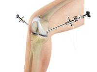
Arthroscopy of the Knee Joint
Knee Arthroscopy is a common surgical procedure performed using an arthroscope, a viewing instrument, to look into the knee joint to diagnose or treat a knee problem. It is a relatively safe procedure and a majority of the patient’s discharge from the hospital on the same day of surgery.
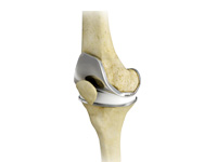
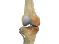
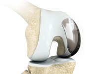
Total Knee Replacement (TKR)
Total knee replacement, also called total knee arthroplasty, is a surgical procedure in which the worn out or damaged surfaces of the knee joint are removed and replaced with artificial parts. The knee is made up of the femur (thigh bone), the tibia (shin bone), and patella (kneecap). The meniscus, the soft cartilage between the femur and tibia, serves as a cushion and helps absorb shock during motion.
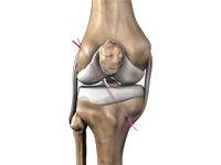
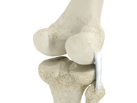
Anterior Cruciate Ligament ACL Reconstruction
The anterior cruciate ligament is one of the major stabilizing ligaments in the knee. It is a strong rope like structure located in the center of the knee running from the femur to the tibia. When this ligament tears unfortunately, it does not heal and often leads to the feeling of instability in the knee.
ACL Reconstruction Hamstring Tendon
Anterior cruciate ligament (ACL) reconstruction hamstring method is a surgical procedure that replaces the injured ACL with a hamstring tendon. Anterior cruciate ligament is one of the four major ligaments of the knee that connects the femur (thigh bone) to the tibia (shin bone) and helps stabilize your knee joint.
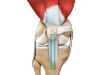
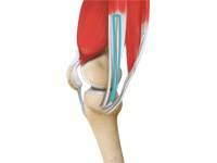
ACL Reconstruction Patellar Tendon
Anterior cruciate ligament (ACL) reconstruction patellar tendon is a surgical procedure that replaces the injured ACL with a patellar tendon. Anterior cruciate ligament is one of the four major ligaments of the knee that connects the femur (thigh bone) to the tibia (shin bone) and helps stabilize the knee joint.
Uni Condylar Knee Replacement
Unicompartmental knee replacement is a minimally invasive surgery in which only the damaged compartment of the knee is replaced with an implant. It is also called a partial knee replacement. The knee can be divided into three compartments: patellofemoral, the compartment in front of the knee between the knee cap and thigh bone, medial compartment, on the inside portion of the knee, and lateral compartment which is the area on the outside portion of the knee joint.
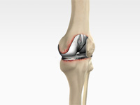
Revision Knee Replacement
Revision knee replacement surgery involves replacing part or all of your previous knee prosthesis with a new prosthesis. Although total knee replacement surgery is successful, sometimes the procedure can fail due to various reasons and require a second revision surgery.